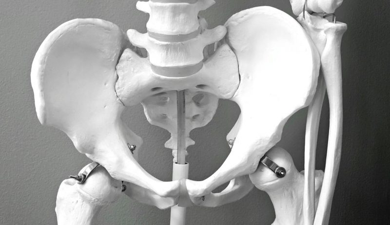
Although all vertebrates possess a pelvis, only humans have evolved a structure optimized for efficient, long-distance bipedal locomotion. This evolutionary transformation took over five million years, yet the precise developmental and genetic mechanisms have long remained unclear.
A recent study published in Nature sheds light on this question by comparing pelvic development during embryonic stages in humans and other mammals. Led by a team from Harvard University, the research analyzed samples from humans, mice, chimpanzees, and gibbons, combining anatomical observations with genomic data, with particular focus on the ilium—the bone that connects the spine and lower limbs and stabilizes gait.
The study identified two critical phases in human pelvic development. Around the seventh week of embryonic growth, the initially vertical cartilage of the ilium undergoes a 90-degree rotation, shortening and widening the pelvis into a bowl-like shape that supports upright walking. The second phase occurs around the 24th week of gestation, when ossification of the ilium is delayed compared with other primates. This prolonged plasticity enables the pelvis to adapt into a structure suited not only for bipedal locomotion but also for childbirth involving large-brained infants.
Researchers also pinpointed five key genes that regulate cartilage growth and ossification, guiding the formation of the pelvis at the molecular level. These findings not only provide a developmental explanation of how humans "stood up", but also highlight the central role of genetic regulation in shaping evolutionary morphology.
Genetic and Developmental Insights Into the Evolution of the Human Pelvis
Tags
A large-scale genetic study published in Nature Medicine has uncovered a surprising paradox: a gene mutation known to cause severe obesity appears to significantly reduce the risk of heart disease…

A 97-million-year-old sea turtle fossil unearthed in Lebanon is reshaping scientists' understanding of how turtles adapted to life in the ocean. The study, published in iScience, a journal under Cell,…

Humans can distinguish up to a trillion different odors—yet this remarkable ability relies on only about 400 types of olfactory receptor proteins. Because these receptors are notoriously difficult to express…

Global conservation groups, scientists and beekeepers are sharply divided over whether to ban the deliberate release of genetically engineered organisms into the wild. Dozens of NGOs have urged the International…

Single-Cell Sequencing Maps Plant Stem Cell Networks, Opening Path to Higher Crop Yields
Plant stem cells are the foundation of agriculture, underpinning the production of food, feed, and biofuels. Yet the core genetic networks controlling their activity have remained poorly understood. A research…

Archaeologists have uncovered stone tools at the Little Paxton site, about 80 kilometers east of London, dating back roughly 440,000 years. Using advanced infrared radiofluorescence dating, researchers confirmed the artifacts…